
Technology & Innovation
BIOMEDICAL ENGINEERING
Electrochemical Catheter Hub Could Prevent Bloodstream Infections

Licensed via Adobe Stock
June 25, 2025
Washington State University and Mayo Clinic researchers have developed an electrochemical catheter hub that could someday help prevent deadly central line-associated bloodstream infections (CLABSIs) that annually kill thousands of people around the world.
Reporting in the journal Biotechnology and Bioengineering, the researchers showed that their e-catheter hub showed “significant antimicrobial activity,” killing off a common type of bacteria that is responsible for hospital infections.
“Our idea was to produce hypochlorous acid, a common disinfectant, at a very low concentration to prevent CLABSI. It eliminated infection,” said Haluk Beyenal, a professor in the Gene and Linda Voiland School of Chemical Engineering and Bioengineering and corresponding author on the paper.
Central venous catheters, flexible tubing that are used in many medical procedures to administer fluids or medications, are responsible for about 20 percent of bloodstream infections.
While hospitals often try to prevent the infections with careful protocols and preemptive antibiotics, the catheters can still be a breeding ground for bacteria, including varieties that are antibiotic-resistant. People handling catheters can easily introduce pathogens when they are connected or disconnected at their hubs, and the danger of infection increases for patients who have a prolonged catheterization. Bloodstream infections can become serious, leading to sepsis which causes thousands of deaths annually and are one of the top causes of death in several North American and European countries.
“During COVID-19, CLABSI rates rose by more than 50% and CLABSIs were significant contributor to increased mortality. In other words, some of the mortalities were due to CLABSI,” said Beyenal.
The WSU researchers have been working for several years to use electric current to create a continual disinfectant to prevent and treat infections.
“The key challenge was whether we could make it usable and if we could control it,” said Beyenal. “We needed to generate a low concentration without generating toxicity but eradicate pathogens.”
The researchers used 3D printing to create a catheter hub, then incorporated small, battery-powered wearable electronics that control electrodes to produce an electrochemical reaction. The electrodes are tiny wires made of gold or titanium. When controlled electric power is added to the electrodes, a reaction occurs, converting salt water in the catheter hub to hypochlorous acid, the bacteria-killing disinfectant.
“When you use a catheter hub system, you always have to fill it up with a solution that generally includes sodium chloride (salt),” said Beyenal. “We simply use the chloride in the sodium chloride.”
Hypochlorous acid is commonly used in cleaning products, including in some hand sanitizers. It is also produced naturally by white blood cells of healthy people to fight bacteria, fungi, and viruses. Instead of one application that one might use with a hand sanitizer, the e-catheter hub is able to continually produce the disinfectant.
The researchers showed the e-catheter hub killed one variety of a common bacteria that cause blood infections. The gold-based electrodes worked best for maintaining steady production of hypochlorous acid and continual killing of bacteria.
“We chose that type of bacteria (to test) because of its resistance to multiple antibiotics, its widespread presence in central venous catheters, and its ability to persist on hospital surfaces and equipment for extended periods,” said Majid Al-Qurahi, first author on the paper and a graduate student in the Voiland School.
The researchers will soon be testing the catheter hub in animals and with other varieties of bacterial infection and at higher levels. They also are investigating the maximum concentration of disinfectant that they can produce to kill off bacteria without causing any bodily harm.
“The catheter is like a direct gateway to the bloodstream, so we need to make sure everything is safe,” said Al-qurahi.
Source: Washington State University
BIOMEDICAL ENGINEERING
Scientists Develop Mask Filter That Blocks Nanoparticles Without Restricting Airflow

Researchers at the University of Tokyo have developed a filter that effectively captures small particles without restricting air flow. Courtesy of Institute of Industrial Science, University of Tokyo
May 21, 2025
When the mesh size of mask fabrics is small enough to capture viruses, which are usually around 100 nanometers in size, the fabric typically also restricts airflow, resulting in user discomfort. But now, researchers from Japan have found a way to avoid this.
In a study published this month in Materials Advances, researchers from the Institute of Industrial Science, the University of Tokyo have overcome this bottleneck and developed a filter capable of capturing nanoparticles such as viruses without greatly restricting airflow. They managed this feat through careful design of the pore structure in the filter.
The filter is constructed from nanosheets consisting of an ordered mesh composed of porphyrins, which are flat, ring-shaped molecules with a central hole. The tiny holes in the porphyrin molecules are suitably sized to allow the easy passage of the small gas molecules in air while blocking the movement of larger particles, such as viruses. The nanosheets are then supported on a fabric modified with nanofibers containing pores of several hundred nanometers to form the filter.
“The porphyrin-based nanosheets are constructed through interfacial reactions that are driven by the movement of reactants caused by the gradient of surface tension at the air–solvent interface, known as the Marangoni effect,” says senior author Kazuyuki Ishii. “The nanosheets are then compressed and coated on nanofiber-modified fabric using a stamp method.”
The team tested their filter using the standard procedure used to test N95 face masks. The results of the particle filtration tests revealed that the filter effectively trapped particles that were as small as viruses. The filter achieved a particle filtration efficiency of 96%, which exceeds the requirement of 95% for an N95 face mask.
“Our porphyrin-based filter collected nanoparticles with a diameter as small as 100 nanometers,” explains Ishii. “Importantly, the filter also showed minimal decrease of differential pressure in gas flow measurements. This indicates that the filter is capable of trapping particles as small as viruses, while barely restricting air flow.”
The team’s approach involving coating porous nanosheets on nanofibers is promising to provide materials capable of effectively filtering small particles like viruses while maintaining air flow to ensure both user comfort and protection.
The article “Hybridization of Nanofiber-modified Fabrics with Porphyrin-based Nanosheets for Nanoparticle Capture” was published in Materials Advances at DOI:10.1039/D5MA00058K.
Source: Institute of Industrial Science, the University of Tokyo
BIOMEDICAL ENGINEERING
AI-Driven Smart Devices to Transform Healthcare, Study Suggests
Courtesy of ChayTee, licensed via Adobe Stock
April 2, 2025
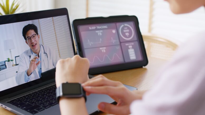
AI-powered, internet-connected medical devices have the potential to revolutionize healthcare by enabling early disease detection, real-time patient monitoring, and personalized treatments, a new study suggests.
They are already saving lives. Wearable devices can detect cardiac issues early, triggering emergency responses and preventing complications.
“The Internet of Medical Things (IoMT), powered by AI, connects medical devices for real-time monitoring and analysis – anything from a smartwatch to hospital monitors. For example, a wearable heart monitor can detect irregular rhythms and send alerts instantly,” said professor of data science Amir Gandomi, from the University of Technology Sydney (UTS). “This study provides a comprehensive roadmap for integrating IoMT into healthcare, showing its potential to improve efficiency and reduce costs, while addressing key challenges like security and the ability to operate with other systems. For patients, it means better health management and fewer hospital visits. For families, it offers peace of mind, especially for seniors or those with chronic conditions. IoMT is making healthcare smarter, safer, and more responsive,” he said.
Gandomi is among the world’s most cited researchers for his work in artificial intelligence and data analytics. He has received multiple prestigious international awards, including most recently the 2024 IEEE TCSC Award for Excellence in Scalable Computing (MCR) and the 2025 Frederick Palmer Prize.
While his research covers a wide range of real-world engineering problems, he has a particular focus on the use of AI and data analytics to improve healthcare, including pandemic response and detection of diseases such as Parkinson’s, diabetes, cancer and heart disease.
The study, Transformative impacts of the internet of medical things on modern healthcare, was led by associate professor Shams Forruque Ahmed from Sunway University in Malaysia, together with Gandomi and an international research team.
“Our research highlights IoMT’s potential to improve patient outcomes, reduce hospital strain, and reduce costs, making it essential for future healthcare systems,” said Gandomi.
It explores IoMT’s full impact on healthcare – its benefits, challenges, and real-world applications, and highlights breakthrough results, such as AI-powered IoMT achieving 99.84% accuracy in heart disease diagnosis from medical imaging, and real-time seizure detection.
The study also examines the challenges of integrating AI-powered IoMT technology into healthcare systems, including the need for strong data security, device compatibility, and better regulations to ensure patient trust and safety.
“For healthcare providers, investing in IoMT means upgrading digital infrastructure, training staff, and adopting remote monitoring for proactive care. IoMT also requires clear regulations and standards to ensure security and patient privacy,” said Ahmed.
Source: University of Technology Sydney
DIAGNOSTICS
Prototype Breath Tests Identify Bacterial Infections in Minutes
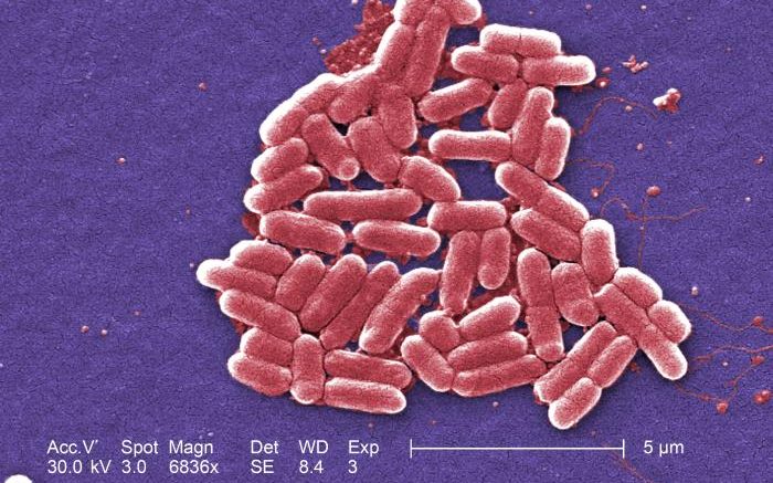
E. coli bacteria. Courtesy of CDC
March 18, 2026
Infectious diseases are a major cause of death worldwide, and diagnosing bacterial infections remains a challenge in medicine. And doing so reliably is more important than ever, given the increasing frequency of antibiotic resistance. Now, research published in ACS Central Science could help healthcare professionals non-invasively diagnose bacterial infections, using breath-based tests. Initial experiments demonstrated the approach in animals with pneumonia and infections in the bloodstream, muscles and bones.
“In designing this study, we were motivated by a developing trend in clinical practice, whereby patients and providers want answers right away that will inform treatment decisions,” says David Wilson, a corresponding author of the study. “If a patient visits the emergency room or clinic, we hope that he or she can be diagnosed with an acute bacterial infection as efficiently as possible.”
Doctors currently rely on blood tests, imaging, cultures and molecular diagnostics to identify the cause of infections, but these tools are limited because they are slow, non-specific or expensive. The start to a potential solution could be the long-used breath test for Helicobacter pylori, a bacterium that causes a common stomach infection. The original test works when a person drinks a liquid containing traceable substances metabolized by H. pylori. Then the person exhales into a device that measures labeled carbon dioxide in their breath, indicating the infection is present. Inspired by this test, Wilson, Kiel Neumann, Marina López-Álvarez and colleagues set out to expand the technology’s capabilities to detect a broader range of bacterial infections.
For their prototype, the team tested sugar and sugar alcohols tagged with carbon-13, a traceable form of carbon that bacteria metabolize but human cells largely ignore. In lab experiments, the researchers identified several of these compounds that bacteria convert into carbon-13-labeled carbon dioxide. Then they analyzed the labeled gas using a simple technique called nondispersive infrared spectroscopy.
When mice with infections such as pneumonia and bone, muscle, or blood infections received intravenous injections of these tagged compounds, the animals’ breath quickly showed elevated levels of the labeled carbon dioxide. Although the breath testing protocol was not optimized in this study, the researchers say they typically saw elevated carbon-13-labeled breath signals in infected animals within the first 10 minutes of metabolite administration and breath sampling. In contrast, the breath of healthy mice showed little to no carbon-13.
In one infection model for E. coli, the amount of labeled carbon dioxide in the breath decreased during antibiotic treatment as bacterial levels went down, suggesting the method could also be used to monitor how well treatments are working.
Because breath-test instruments are portable and breath signals appear within minutes after the traceable carbon-13 is administered, the test could potentially deliver results faster than current methods. In addition, the sugar and sugar alcohols used are considered safe for humans, and the researchers say this approach could eventually become a tool for diagnosing bacterial infections.
The authors have filed a U.S. patent related to this work.
Source: American Chemical Society (ACS)
BIOMEDICAL ENGINEERING
Nurse-Engineer Team Honored for Inventing IV Pole Designed to Improve Patient Safety
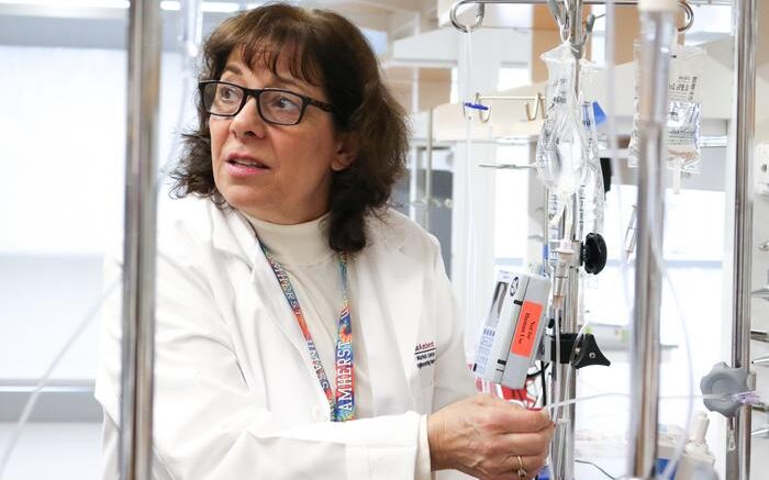
Karen Giuliano, nursing co-director of the Elaine Marieb Center for Nursing and Engineering Innovation, conducts research in the center’s IV lab. Courtesy of Lauren LeCours/UMass Amherst
March 12, 2025
A nurse-engineer team at the University of Massachusetts Amherst has been honored with an ANA Innovation Award for inventing a new intravenous (IV) pole designed to improve the safety and ease of administering IV medications at the hospital bedside.
The American Nurses Association Foundation and the American Nurses Enterprise announced the 2025 award winners on March 11. The team – Karen Giuliano, nursing co-director of the Elaine Marieb Center for Nursing and Engineering Innovation; Jeannine Blake, assistant professor of nursing; and Juan Jiménez, associate professor of mechanical and industrial engineering and a Manning/IALS Innovation Fellow – won honorable mention in the 2025 Team Innovation Award category.
Preventable medication errors harm approximately 500,000 hospitalized patients in the U.S. each year. Many of these errors occur with the use of IV smart pumps, which require a very specific system setup to ensure the right amount of medication is delivered as ordered. When the setup is not followed, the IV smart pump can deliver too much or too little of the medication prescribed, even as the pump signals it is delivering the correct amount.
By combining the engineering and fluid dynamics expertise of Jiménez with the clinical knowledge of critical-care nurses Giuliano and Blake, this interdisciplinary team set out to develop an IV pole that simplifies and accelerates the setup and delivery of IV medications in hospitals when using an IV pump, ultimately reducing the occurrence of dangerous yet largely preventable medication errors. The idea stemmed from real-world clinical observations, which revealed that standard IV poles often made it more challenging and time-consuming for frontline nurses to achieve optimal IV infusion setups.
A patent is under review for this novel IV pole, which features an adjustable crossbar for hanging infusions. This innovative crossbar automatically maintains the required height differential between the IV pump and the medication container. Established by IV smart pump manufacturers, this differential helps ensure optimal fluid flow accuracy. The pole improves IV medication delivery efficiency while minimizing the need for manual adjustments.
“The work of Drs. Giuliano, Blake and Jiménez, along with the Elaine Marieb Center, represents the future of healthcare innovation,” said Frank Sup, engineering co-director of the Elaine Marieb Center.
Giuliano, Jiménez and Blake received a 2022 Manning/IALS Innovation Award to support work on their new IV pole project. “By bringing together expertise from both nursing and engineering, we are breaking barriers and reimagining how technology can support nurses and improve patient care,” Giuliano said.
According to Jiménez, “This project is a perfect example of why engineering and nursing must work together to solve real-world healthcare challenges. Nurses bring firsthand clinical experience and deep knowledge of patient care, while engineers contribute technical expertise to design practical, effective solutions.”
The ANA Foundation’s Team Innovation Award celebrates interdisciplinary collaboration and ingenuity in addressing critical healthcare challenges. The winning teams exemplify these values by integrating engineering principles into nursing practice to develop cutting-edge healthcare solutions that enhance clinical outcomes and streamline nursing workflows. Their efforts have contributed to novel medical devices and improved patient safety protocols, according to the ANA Foundation.
The invention of the new IV pole is part of the Elaine Marieb Center for Nursing and Engineering Innovation’s active program of research on the safety and usability of IV smart pumps. Their research has focused on reducing infusion errors, optimizing alarm management and enhancing usability to better support clinicians in high-pressure environments, such as the intensive care unit (ICU), where patients are typically receiving multiple IV drips at the same time.
Source: University of Massachusetts Amherst