

Pathogens
Evolutionary History May Explain Why Some People Develop More Severe COVID-19 Than Others
Aug. 20, 2026
A study led by researchers at the USC Dornsife College of Letters, Arts and Sciences and Howard University suggests that some of the genes involved in the body’s immune response today bear the marks of ancient battles with infectious diseases.
The research also identified rare genetic variants that may help explain why some people developed more severe COVID-19 than others. The findings connect two fields that are rarely studied together: human evolution and infectious disease. By bringing them together, the scientists uncovered clues that may have been missed by studying either field alone.
The study traced the evolutionary history of four immune system genes, which were previously linked to respiratory or inflammatory diseases, across populations around the world. The research team then examined whether changes in those genes were linked to severe COVID-19 in nearly 4,000 people in Italy, using data from the Italian GEN-COVID study.
For Michael Campbell, associate professor of biological sciences at USC Dornsife and the study’s senior author, understanding how evolution shaped the immune system over thousands of years offers a new way to think about why people respond differently to disease today.
“SARS-CoV-2, the coronavirus that causes COVID-19, may be new, but the immune system it encountered isn’t,” Campbell said. “Our immune genes have been shaped by thousands of generations of encounters with pathogens. Understanding the evolutionary history of immune genes can provide insights into why people respond differently to infectious diseases today.”
How have ancient diseases shaped the human immune system?
To explore that history, the scientists at USC Dornsife and Howard collaborated with colleagues at Yale and Columbia universities to analyze four genes that help regulate the body’s immune response — IL-4, TLR2, CCL2 and SLC11A1. Using genetic data from more than 2,000 people representing populations from Africa, Europe and Asia, they searched for signs that evolution had favored particular versions of these genes over time, leaving lasting marks in the human genome.
The team found evidence that all four genes had been shaped by natural selection, but not in the same way everywhere. Different versions of the genes were more common in different populations, suggesting infectious diseases and other environmental pressures may have shaped human evolution differently around the world.
Some of those gene variants were also found in the DNA of Neanderthals or Denisovans, extinct human relatives that lived tens of thousands of years ago. In some cases, the similarities likely reflect inherited DNA from a shared ancestor. In others, they point to ancient interbreeding between early modern humans and these extinct relatives.
Importantly, Campbell said, these evolutionary changes almost certainly happened long before the coronavirus emerged. “The signals we found weren’t created by COVID-19,” he said. “They reflect much older evolutionary pressures, probably from infectious diseases our ancestors faced over thousands of years.”
Rare gene variants linked to severe COVID-19
The researchers then turned to a different question: Could some of these immune genes also help explain severe COVID-19?
Using genetic and clinical information from nearly 4,000 people in Italy who tested positive for COVID-19, the researchers identified two rare changes in the TLR2 gene. One was found more often in COVID-19 patients who had previously received an organ transplant than in other COVID-19 patients. The second was associated with more severe forms of COVID-19, suggesting it may contribute to differences in how seriously ill some people become.
Unlike the common gene variants shaped by evolution over thousands of years, these TLR2 variants were rare and appear to be much more recent genetic mutations.
Because organ transplant recipients are already known to face a higher risk of severe COVID-19, the finding suggests a TLR2 variant could be one factor influencing that increased risk. The researchers say, however, that more research is needed to determine if that’s the case.
The researchers also caution that the COVID-19 findings overall should be interpreted carefully. The genetic associations will need to be confirmed in larger, independent populations and validated through laboratory studies. They also note the need to include more genetically diverse populations in future studies to better understand disease risk worldwide.
Why evolution still matters today
By identifying immune genes shaped by ancient infectious diseases, such as TLR2, scientists may gain new clues about the biological basis of infectious disease susceptibility.
“The pathogens our ancestors encountered thousands of years ago helped shape the immune system we carry today,” Campbell said. “By tracing that evolutionary history, we can better understand why people respond differently to infectious diseases today and identify biological pathways that may inform new strategies for disease prevention and treatment.”
In addition to Campbell, study authors include Alessandro Lisi and Marisol Fermin Flores of USC Dornsife; Thomas Heinbockel and Kareem Washington of Howard University; Christopher N. Cross of Howard University and Yale University; and Faith C. Simmonds of Columbia University.
Source: University of Southern California
Severe COVID-19 Reactivates Dormant Viruses, Study Finds
Aug. 20, 2026
Better understanding of how these activated viruses contribute to COVID-related outcomes could help physicians better predict potential complications and improve treatment for patients with severe COVID infection
Chronically infecting viruses — such as Epstein Barr, cytomegalovirus (CMV), and herpes virus — are common, and often innocuous and asymptomatic. However, emerging evidence suggests their reactivation may contribute to autoimmune disease and other chronic conditions. In a new Nature study involving 15 biomedical research institutions across the United States, Boston Children’s Hospital researchers and their collaborators have discovered that COVID-19 reactivates certain dormant viruses in hospitalized patients. These findings expand understanding of chronically infecting viruses and could inform development of strategies to combat their reactivation.
Physician scientist Ofer Levy, MD, PhD, director of the Precision Vaccines Program (PVP) at Boston Children’s, served as a site principal investigator for this National Institutes of Health (NIH)-funded study of 1,154 patients across 20 U.S. biomedical research hospitals that was designed to define biomarkers of COVID severity and outcomes. The research team used genomic sequencing to look for reactivated viruses in the patients since long-cleared viral infections can sometimes reawaken in times of stress.
“This is the largest and most comprehensive biomarker study of COVID-19, in which we followed more than 1,000 patients, collected more than 200,000 samples, and generated more than 1 billion data points over the course of a year for this public resource,” says Joann Diray Arce, PhD, who leads the PVP-Data Management and Analysis Core and is the lead of the study’s Clinical and Data Coordinating Center.
The research team detected 11 reactivated viruses in patients within the first 40 days from admission, with the most detected ones being Epstein-Barr, herpes simplex 1, cytomegalovirus, and Anelloviridae viruses. Notably, reactivation of Anelloviridae, a poorly understood family of viruses typically latent in about 90 percent of the population, was associated prominently with long-term physical disability and long COVID.
“This association with long COVID is an interesting finding as millions around the world suffer from this chronic condition,” says Levy. “Having new insight as to the molecular and viral associations with long COVID could point the way to better understanding and ultimately better diagnostics and treatments.”
In an analysis of the blood samples from the patients, Epstein-Barr and cytomegalovirus seemed to activate in response to inflammation rather than immune system suppression. The researchers say this is a surprising new mechanism, challenging the prevailing view that chronic viral reactivation is primarily a consequence of immunosuppression. This finding demonstrates that reactivations occur frequently in apparently immunocompetent individuals during severe illness and in association with increased systemic inflammation.
“Although many no longer think of COVID being a problem, up to 50,000 Americans died of COVID in 2025-2026 respiratory season and some estimates suggest over 10 million U.S. adults suffer from long COVID,” says Levy. “We need to help these patients recover with the best outcomes.” He adds “Moreover, sooner or later, there may be another coronavirus pandemic, which means we need to learn all the lessons we can from COVID-19 to be better prepared.”
Next steps for this work will be to uncover how the immune system responds to these viruses over the course COVID-19, with the aim of identifying effective therapeutics and establishing the optimal timing of any interventions.
Source: Boston Children’s Hospital
The First 24 Hours After Viral Infection are Critical, Study Finds
Courtesy of Riho Saito
Aug. 20, 2026
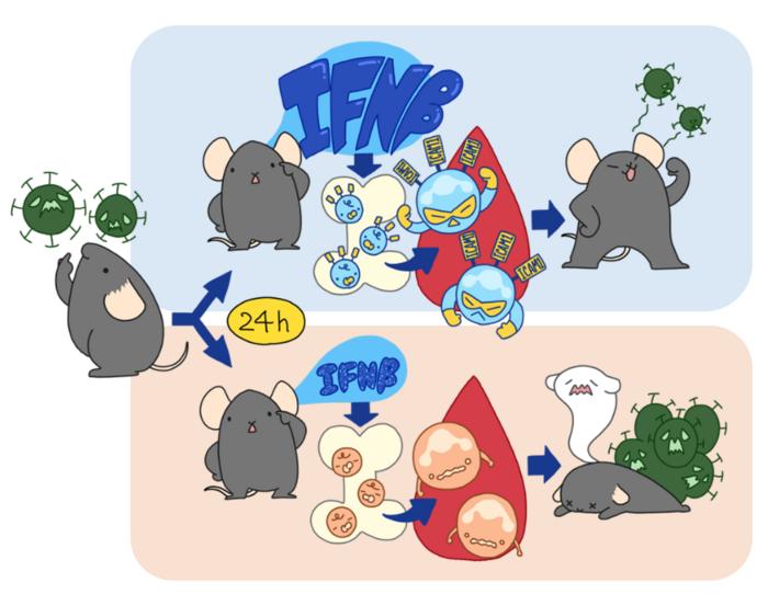
The first 24 hours after a viral infection are critical and can help determine whether a mouse will survive. The outcome depends on how its immune system responds to the virus, particularly through a molecule called type I interferon (IFN-β).
Age, genetics and lifestyle are known to influence how people respond to viral infections. Scientists also know that differences in immune response play an important role, but exactly how these factors shape the course of an infection has remained unclear. Now, new research from Hokkaido University, using mouse models, suggests that a critical part of the answer may lie in how the immune system responds during the first 24 hours after infection.
Published in iScience, the study shows that, in mice, a powerful early immune response can determine whether an individual survives a severe viral infection. The researchers identified a previously unrecognized immune “checkpoint” that shapes the body’s antiviral defenses and could guide the development of future treatments for severe viral diseases.
Using genetically identical mice raised in the same controlled environment, the researchers infected the animals with a lethal virus called vesicular stomatitis virus (VSV). Some mice survived while others did not.
The team discovered that the surviving mice released a rapid burst of type I interferon (IFNβ), an immune signaling protein that helps coordinate the body’s defense against viruses. Mice that generated this early interferon response were much more likely to survive.
The early interferon response triggered another group of immune cells called neutrophils. Specifically, it induced a distinct population of neutrophils expressing the cell-surface marker ICAM1, which exhibited heightened inflammatory signaling and an enhanced antiviral immune profile.
When the researchers blocked type I interferon during the first 24 hours after infection, most of the mice died. But, blocking it two days after infection had little effect.
The findings suggest that there is a critical window early in infection during which the immune system decides to establish an effective antiviral response.
“Biological variability should not always be viewed as experimental noise,” says lead author of the study Associate Professor Tomohiko Okazaki. “Instead of treating differences between genetically identical individuals as experimental noise, we viewed them as a source of biological insight. That approach allowed us to uncover an early immune checkpoint that would have been difficult to identify using conventional comparisons.”
The findings indicate that treatments designed to strengthen or mimic this early immune response could improve outcomes in severe viral infections. Although the study was conducted in mice and further research is needed to determine whether a similar mechanism operates in humans, the work provides a clue into why identical infections can produce dramatically different outcomes.
“Since the COVID-19 pandemic, there has been growing interest in understanding why viral infections can produce dramatically different outcomes among individuals,” Okazaki said. “Age and underlying medical conditions are well-known risk factors, but they do not fully explain this variability. Our findings identify an early immune mechanism that may help account for these differences and could inform future therapeutic strategies.”
Source: Hokkaido University
Hepatitis E Viruses Mutate Early During Infection
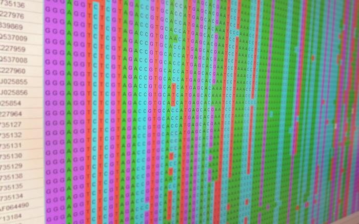
The researchers analyzed the virus’s genetic information during the acute phase of infection. Courtesy of Daniel Todt
July 13, 2026
Otherwise healthy individuals usually do not notice an acute hepatitis E infection; the immune system generally eliminates the viruses within a few weeks. However, the infection can become chronic in individuals with a compromised or medicinally suppressed immune system.
In order to better understand which changes occur during the initial weeks of the infection, the research team specifically focused on this early phase. “The virus develops during an acute infection,” explains Saskia Janshoff, doctoral student and first author of the study. “We wanted to learn how diverse the virus populations are during this period and which changes occur frequently.” The analyses showed relatively low genetic virus diversity among the blood donors compared to the chronic phase of the infection. However, the researchers were able to identify certain changes that recurred in multiple donors.
For sequencing, the team focused on the viral polymerase in particular. This enzyme is important for the replication of the virus and is also a target for antiviral medications. The researchers discovered four sites in the genetic material where similar changes occurred especially frequently.
In lab experiments, the researchers examined the effects of these mutations. “We noticed that some variants of the virus were barely capable of replicating on their own,” says Dr. André Gömer. Nevertheless, they could apparently exist within mixed virus populations. This is possible through a mechanism known in the field as transcomplementation: Defective virus variants benefit from the simultaneous presence of intact viral polymerases and can thus still be replicated.
Various samples from individual blood donors who were examined repeatedly throughout the course of infection provided further insight. Changes in the makeup of the virus population in individual patients within a few weeks was observed. “The early stages of an infection are highly dynamic,” says Gömer. “Individual variants occur, alter their frequency, or disappear. Such processes can only be made visible by repeatedly taking samples.”
It has not yet been fully explained why some mutations occur and exist temporarily despite their limited replicability. The researchers assume that certain changes could influence the interaction with the immune system. However, further studies are required to show whether this actually benefits the viruses. The study thus provides new insight into the early evolution of the hepatitis E virus and lays a foundation for future work on the significance of genetic changes for the course of the disease and the response to therapy.
Researchers from Ruhr University Bochum, the Ruhr University Bochum University Hospital, University of Bielefeld, the HepE-Hub, TWINCORE Hannover, the Hannover Medical School, and the German Center for Infection Research were involved in the work.
Source: Ruhr University Bochum
Researchers Find a Common Weakness in Major Gut Pathogens
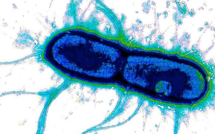
WashU Medicine researchers and collaborators at the University of Missouri identified a shared vulnerability across diarrhea-causing bacteria including E. coli (shown) and Shigella, a finding that could potentially lead to a single combination vaccine against these pathogens. Courtesy of David Hasty
June 18, 2026
The bacteria enterotoxigenic E. coli and Shigella together cause hundreds of millions of infections each year and are among the leading causes of diarrheal death, especially in children. Decades of vaccine development efforts have come up short, in part because the usual vaccine targets vary too much from one strain to the next.
New research from Washington University School of Medicine in St. Louis points to a shared biological feature of these gut pathogens that could lead to a vaccine that protects against both.
Researchers at WashU Medicine, along with collaborators at the University of Missouri and the International Centre for Diarrheal Disease Research in Bangladesh, found that enterotoxigenic E. coli (the leading cause of travelers’ diarrhea), Shigella and other diarrhea-causing pathogens rely on three closely related enzymes to get through the gut’s protective mucus layer and cause infection. Based on samples from infected patients and volunteers exposed to the bugs, the team showed that antibodies targeting one shared region of these enzymes can neutralize all three biomolecules and block the bacteria from penetrating the mucus barrier of the intestines.
The results, which appear June 15 in PNAS, point to the potential for a single combination vaccine against these major causes of severe diarrhea.
“For something so common and so deadly to young children, it’s striking that we still don’t have a vaccine for either of these pathogens,” said James M. Fleckenstein, MD, a professor of medicine in the Division of Infectious Diseases at WashU Medicine and co-senior author on the study. “What’s exciting here is that we’ve found a kind of Achilles’ heel or weak point they share that we might be able to target to protect against both.”
To cause illness, gut pathogens must first break through a thick layer of mucus that coats the intestine and holds even the body’s healthy resident bacteria at bay. Getting past that barrier is a critical early step in infection — and a point where, Fleckenstein said, harmful bacteria might be stopped without disrupting beneficial microorganisms. Enterotoxigenic E. coli (ETEC) — so named because it causes gastrointestinal disease, unlike other strains of E. coli that are harmless — and Shigella manage the task using closely related enzymes that cut through the main protein in gut mucus. Once they breach the barrier, the bacteria can deliver the toxins that cause diarrhea.
Fleckenstein’s lab first identified one such enzyme in diarrhea-causing E. coli, called EatA, which fittingly eats away at the primary structural component of mucus. The team has now shown that two related enzymes — SepA and Pic, produced by Shigella and some other diarrhea-causing bacteria — perform the same mucus-busting function.
Working with coauthor Ali Ellebedy, PhD, the Leo Loeb Professor in the WashU Medicine Department of Pathology & Immunology, Fleckenstein and collaborators isolated antibodies from patients in Bangladesh naturally infected with ETEC and from volunteers intentionally infected with the bacteria in controlled studies. They found that antibodies blocking EatA also neutralized SepA and Pic. Antibodies are proteins the immune system produces to recognize a specific target and lock onto it so that it can be destroyed.
Structural biologists at the University of Missouri, including first author David P. Buckley, PhD, a postdoctoral research associate, then used cryo-electron microscopy — a technique that flash-freezes molecules to image them in fine detail — to pinpoint exactly where the most effective antibodies latched onto the enzymes. The spot turned out to be a region shared across all three, which explains how a single antibody can disable the mucus-degrading machinery of multiple pathogens. It also gives vaccine designers a precise target for generating a vaccine that would prompt the immune system to produce such antibodies and have them ready in case of infection.
“This study establishes EatA as a viable vaccine candidate capable of providing protection across multiple pathogens,” said Zachary Berndsen, PhD, an assistant professor of biochemistry at the University of Missouri and co-senior author on the study. “By identifying the key regions of EatA that are targeted by neutralizing antibodies capable of inhibiting its enzymatic function, we’ve established a foundation for rational vaccine design — a major advance toward development of effective therapeutics that have the potential to save many lives.”
The project builds on earlier studies of children in Dhaka, Bangladesh, showing that those who naturally develop antibodies against EatA tend to be protected from illness, while children without them are more likely to get sick.
The need for vaccines to protect against these infections isn’t confined to the developing world. Enterotoxigenic E. coli has caused large foodborne outbreaks in the United States, and because it is hard to distinguish from harmless E. coli in most clinical labs, cases often go unrecognized. The reliance on antibiotics to treat these infections also fuels antibiotic resistance, which does not respect borders, Fleckenstein noted.
The team is now working to move toward vaccine development.
“These bacteria have evolved right alongside us, and they’ve gotten very good at breaching our defenses,” Fleckenstein said. “If we can block that first step, we have a chance to stop these infections before they ever take hold.”
Buckley DP, Akhtar M, Thapa M, Schmitz A, Turner J, Vickers TJ, Khatoon N, Kaisar MH, Coggin JA, Ganguli D, Sheikh A, Laird RM, Poly F, Porter CK, Ruiz-Perez F, Miller MJ, Chowdhury F, Bhuiyan TR, Qadri F, Trillo-Muyo S, Dolan B, van der Post S, Ellebedy A, Berndsen ZT, Fleckenstein JM. Human enterotoxigenic Escherichia coli (ETEC) infections elicit antibodies that broadly neutralize mucinases of pathogenic Escherichia coli and Shigella. PNAS. June 15, 2026. DOI: https://doi.org/10.1073/pnas.2614012123
This work is supported by the National Institute of Allergy and Infectious Diseases (NIAID) of the National Institutes of Health (NIH), grant numbers R01 AI089894 and R01 AI126887, and by the Department of Veterans Affairs, grant number 5I01BX001469-05. The content is solely the responsibility of the authors and does not necessarily represent the official views of the NIH or the Department of Veterans Affairs.
Source: WashU Medicine
Scientist Leads Research to Break Through Harmful Bacterial Biofilms
UCF College of Medicine assistant professor Renee Fleeman and her lab group work to understand how to fight some of the strongest and most harmful bacteria to humans. Courtesy of UCF College of Medicine/Kadeem Stewart
June 4, 2026
UCF College of Medicine assistant professor Renee Fleeman continues to refine a powerful therapy for drug-resistant bacteria that pierces the gooey coating that anchors and protects such germs from the drugs we take to kill them.
Her research, backed by a five-year $813,130 National Institute of Allergy and Infectious Diseases grant, found that an antimicrobial peptide naturally found in cows weakens the biofilm defenses of Klebsiella pneumoniae bacteria and destroys it.
Now in their fourth year of research, Fleeman and her lab have discovered exactly how the peptide works in findings published in PLOS Pathogens.
“Our research is very advantageous for healthcare because about 80% of bacterial infections being treated in the clinic are bacteria living in a biofilm state, which makes them resistant to virtually every antibiotic available,” she said.
The results represent a critical step to potentially applying this peptide as a therapy and eventually treating patients, as the findings show they can and kill biofilm-embedded bacteria in animal models.
K. pneumoniae is found in the intestines and is usually harmless, however, the bacterium develops resistance over a person’s lifetime as they are exposed to antibiotics. The bacteria also can spread from the intestine to other parts of the body in immunocompromised patients and those who have internal ruptures or exposure to contaminated medical devices. That exposure can lead to pneumonia, urinary tract or wound infections.
“What happens is the bacteria infects the wound, proliferates, and then invades through the bloodstream where it travels to the liver, kidneys and spleen,” Fleeman said. “We found our peptide was able to decrease the bacteria at the source while limiting the bacteria’s ability to move through the blood.”
Fleeman and her lab’s most recent study found that the peptide triggers a dual stress response that tricks the bacteria to break out of their protective biofilm.
They discovered the genetics of a specific protein in the bacterium when turned on in the germ causes it to break from its own protective biofilm. The peptide, in effect, damages the protection and then stresses the bacterium into shedding its protection, making the germ more sensitive to antibiotics and the body’s immune system.
“By hitting the membrane as well as protein synthesis at the same time, it’s a double punch that triggers a genetic change in the cell to make it think it needs to break out of the biofilm as a response to our peptide,” Fleeman says.
The team says their sustained research aims to demonstrate that their peptide can work synergistically with existing antibiotics. They envision long-term applications could involve a topical cream that weakens the bacteria’s defenses and allows standard antibiotics to work more effectively.
“We’re moving our research forward and we’re very hopeful,” Fleeman said.
The first author of this new work is Robert Beckman ’23, who graduated from UCF with a bachelor’s degree in health sciences , managed Fleeman’s lab and is now on his way to the University of Michigan for his Ph.D.
His previous work as an EMT gave him firsthand exposure to infectious diseases and their impact on patients. He says helping to lead the study and working with Fleeman helped prepare him for a career in medical research.
“I have developed a strong foundation in research and gained insight into the many components that define an effective scientist,” he said. “My long-term goal is to remain in academia and eventually lead my own research lab. I plan to continue focusing on bacteriology, with a particular emphasis on pathogenic bacteria and drug discovery applications.”
Source: UCF
Scientists Take Crucial Step in Developing World’s First Measles Treatment
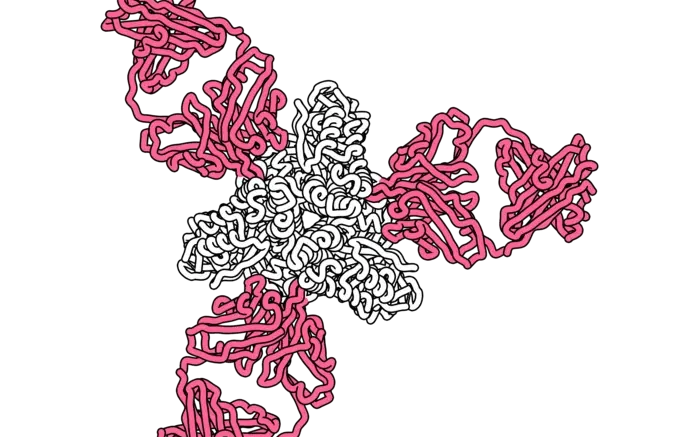
This rendering shows a key measles protein (white) targeted by neutralizing human antibodies (pink). Courtesy of Dawid Zyla, La Jolla Institute for Immunology
May 9, 2026
Scientists at La Jolla Institute for Immunology (LJI) are the first in the world to characterize human antibodies capable of neutralizing measles virus. These antibodies bind to key sites on measles virus and prevent the virus from entering host cells.
The new panel of human antibodies may form the basis for future medical therapies against measles infection. In the study, an infusion of these antibodies resulted in a 500-fold lower viral load in a rodent model of measles infection.
“These antibodies work as prophylaxis—to protect from initial infection—and they work after viral exposure as a treatment to fight measles infection, ” says LJI Professor, President & CEO Erica Ollmann Saphire, PhD, who led the new Cell Host & Microbe study. “It may be possible to give someone an infusion of these antibodies and deliver the immune response they wish they had.”
In recent years, decreased vaccination rates have led to deadly measles outbreaks across the United States and around the world. This sharp rise in measles cases is especially dangerous for the millions of people who cannot receive a measles vaccine.
The measles vaccine is incredibly safe and effective, but it does contain a live, weakened virus. This means people who have compromised immune systems, such as those who are pregnant or receiving chemotherapy, including children, cannot receive the vaccine. The very young are also at risk. Infants must wait until they are 12 months old to be vaccinated, and most children in the United States aren’t fully vaccinated against measles until age 6.
“There are a growing number of people that can’t be vaccinated or haven’t been fully vaccinated,” says Saphire. “The very same people who can’t be vaccinated or can’t be vaccinated yet, are the same people for whom a measles virus infection would be the most severe—or be lethal.”
Until recently, enough people were vaccinated against measles virus that the risk of exposure for this unvaccinated group was very low. Unfortunately, that community protection, called “herd immunity,” is no longer.
LJI scientists are on a mission to find treatment options for the most vulnerable.
Right now, there are no measles-specific therapies to help patients. The new study shows that monoclonal antibody therapies may be the way forward.
Monoclonal antibody therapies work because they contain many copies of a neutralizing antibody. These therapies are widely used for a variety of infectious diseases. Even infants receive monoclonal antibody therapies each year to prevent respiratory syncytial virus (RSV).
To design a monoclonal antibody treatment for measles, researchers need a clear picture of how human antibodies fight the virus.
Saphire and her colleagues began by harnessing an imaging technique called cryo-electron microscopy (cryo-EM) to capture the first-ever glimpses of how antibodies bind to the measles virus. They started by examining mouse antibodies, and they published that work in a recent Nature Communications paper.
That initial study showed—in stunning detail—where measles virus is vulnerable to antibody attack. The mouse antibodies latched onto one key part of the measles virus, called the fusion protein, to block the virus from entering a host cell.
Could human antibodies do the same thing? To find out, the researchers analyzed blood from a clinical research volunteer. This volunteer had been vaccinated against measles many years before, so they already had antibodies ready to fight measles virus.
From this one blood sample, the LJI scientists isolated antibodies that bind to the measles fusion protein, and other antibodies that bind to the second key piece of the virus, an attachment protein called “H.”
They then captured 3D images of these antibodies bound together with the measles virus.
“We found that these antibodies are exceptionally potent,” says LJI Instructor Dawid Zyla, Ph.D., who served as study co-first author. “Two orders of magnitude better than comparable molecules reported at conferences.”
Measles virus is a shape-shifting virus. When it meets a human cell, it unfolds to reveal viral machinery that fuses with the host cell membrane. The new study shows that antibodies targeting the fusion protein work by locking the protein in place, leaving the virus unable to shape shift and infect a host cell.
The next step was to test these antibodies in a preclinical animal model.
Study collaborators at The Ohio State University carried out key experiments using cotton rats as a model. They found that all four lead antibodies reduced the viral load when given either before measles exposure or within 24 to 48 hours after infection. One, an antibody called 3A12, which binds to a site on the F protein, rendered the circulating virus actually undetectable.
While more work needs to be done, the researchers see these antibodies as promising tools in the fight against measles. Their new images of the antibody structures provide the materials needed to make the world’s first before- or after exposure treatment for measles virus.
“Now we know what we’re aiming for, and we have the antibodies we need,” says Saphire.
Additional authors of the study, “Uncovering the features of Measles-targeting human antibodies elicited by the MMR vaccine,” are Marissa Acciani (co-first author), Gele Niemeyer, Stephanie Harkins, Diptiben Parekh, Emily Pawlack, Davide Lacarbonara, Dhvanir Kansara, Margaret E. Ackerman, Stefan Niewiesk, Matteo Porotto, and Kathryn M. Hastie (co-corresponding author).
Source: La Jolla Institute for Immunology
Researchers Crack Genetic Code of Growing Bacterial Threat

Courtesy of CDC
March 31, 2026
Scientists at Houston Methodist Research Institute have discovered that a fast‑rising strep bacterium comes in more forms than expected, including ones that may lead to life-threatening infections.
The study, led by James Musser, MD, PhD, chair of the Department of Pathology and Genomic Medicine and director of the Center for Infectious Diseases at Houston Methodist, is published in Microbiology Spectrum. It is the largest U.S. study investigating Streptococcus dysgalactiae subspecies equisimilis (SDSE) at this level, as these strep infections are increasing worldwide.
The bacterium is most closely related to the flesh-eating Group A Strep, which causes illnesses ranging from mild strep throat and fever to severe conditions like blood infections and necrotizing fasciitis (flesh-eating disease). SDSE was originally considered rare and was believed to mostly infect people who already had other health issues.
First author, Lydia Pouga, PhD, research scientist at Houston Methodist said, by using whole-genome sequencing to analyze more than 800 patient samples, the researchers discovered 44 distinct variants of the bacterium, an insight older testing methods did not provide.
“We discovered that certain strains caused specific types of infection,” Pouga said. “For example, one type of strain was associated with skin infections, another type was associated with blood infections and yet another strain was associated with throat infections. This is the first time we’ve seen such strong associations between strains and specific infections across a large patient population.”
Pouga said the study provides critical genetic data to understand how the bacterium spreads, changes and causes infections. The information will help deliver insights that can improve diagnosis, infection control and future vaccine planning.
Musser’s lab is renowned for using an integrated interdisciplinary research approach to unravel new information about infections caused by group A Streptococcus (GAS), which is responsible for more than 700 million cases of human disease each year globally. Jim Henson, creator of the Muppets, died of a similar form of the disease in 1990.
Other authors include Houston Methodist researchers Stephen Beres, Randall Olsen, Wesley Long and Edward Graviss.
Source: Houston Methodist Research Institute